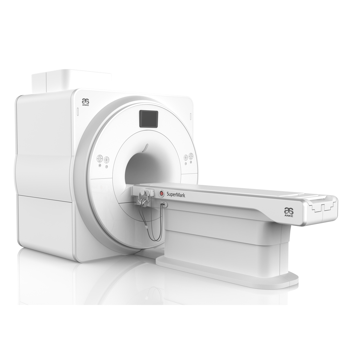
MRI Machine SuperMark 1.5T
Technical Advantages:
- Reliable short bore superconducting magnet system with zero liquid helium
consumption; - New generation fully digitalized and extensible multichannel spectrometer;
- Powerful high efficiency and high fidelity gradient system;
- Multi-channel phased array (PA) RF receiving coil with intelligent identification;
- English operating system and high extensible computer system;
- High resolution conventional clinical images;
- Practical advanced functional imaging.
Superconducting MRI System:
- Highly open and humanization design -> Streamlined design
- Rich sequences and technology satisfy clinical needs -> Efficient service
Low Investment:
- High cost-effective superconducting MRI system;
- Zero liquid helium consumption, low running and maintenance cost;
- Upgrade fully supported by core technologies with patents;
- Low power consumption;
- Compact magnet design, minimum installation space: 35 square meters.
High Return:
- High resolution images with thin slice thickness improve diagnosis;
- Short bore magnet design makes patients comfortable;
- Fast scan speed improves work efficiency.
Technical Specifications:
Magnet system
| Magnet Parameters | |
| Magnet type | Superconducting |
| Magnetic field strength | 1.5T ± 1% |
| Magnetic field stability | ≤ 0.1ppm/h |
| Weight (including 100% liquid helium) | 3800kg |
| Magnet length | 157cm |
| System length (cover to cover) | 170cm |
| Magnet bore diameter | 605mm±5mm |
| Magnet scan bore length | 122cm |
| Shielding method | Active shielding |
| 5 Gaussian fringe field (X, Y, Z) | ≤ 2.6 m × 2.6 m × 4.0m |
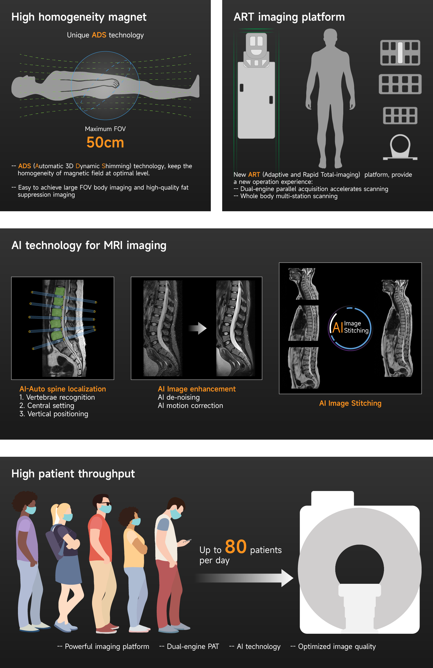
| Shielding methods: Passive Shimming + Active Shimming + 3D Dynamic Shimming | |
| Shimming | |
| Number of superconducting shim coils | 20 |
| Longest time of 3D dynamic shimming | Approx. 30 seconds |
| Magnetic field homogeneity (VRMS measurement method, 24 points 24 sides) | |
| 50 cm DSV (VRMS) | ≤ 0.800ppm |
| 45cm DSV (VRMS) | ≤ 0.450ppm |
| 40cm DSV (VRMS) | ≤ 0.170ppm |
| 30cm DSV (VRMS) | ≤ 0.058ppm |
| 20cm DSV (VRMS) | ≤ 0.020ppm |
| 10cm DSV (VRMS) | ≤ 0.002ppm |
| Magnet cooling system | |
| Provide 4K cold head & Liquid helium "zero" boil-off technology | |
| Liquid helium capacity (100% liquid helium filling) | 700L |
| Liquid helium refilling interval 1) | ≥ 5 years |
1) For typical clinical use, depending on sequences and running state of helium compressor. The system needs to be serviced at regular interval. Undisturbed magnet cooling for 7×24 hours.
Gradient system
| General features | |
| Gradient amplifier type | Ultrafast solid-state technology |
| Gradient amplifier cooling type | Water cooling |
| Gradient coil cooling type | Water cooling |
| Gradient control technology | Digital real-time transmit and receive |
| Gradient performance | |
| Max. amplitude (single axis) | 40mT/m |
| Max. Effective amplitude | 69mT/m |
| Max. slew rate (single axis) | 150T/m/s |
| Max. Effective slew rate | 259T/m/s |
| Min. rise time | 0.27ms |
| Duty cycle | 100% |
| Resolution parameters | |
| Min. 2D thickness | 0.1mm |
| Min. 3D thickness | 0.05mm |
| Max. FOV | 50cm |
| Min. FOV | 1cm |
| Maximum acquisition matrix | 1024×1024 |
| Sequences parameters | |
| FSE shortest TE (256 x 256 matrix) | ≤ 4ms |
| FSE shortest TR (256 x 256 matrix) | ≤ 8ms |
| FSE shortest TE (128 x 128 matrix) | ≤ 3ms |
| FSE shortest TR (128 x 128 matrix) | ≤ 6ms |
| 3D GRE shortest TE (128 x 128 Matrix) | ≤ 0.4ms |
| 3D GRE shortest TR (128 x 128 matrix) | ≤ 1ms |
| 3D GRE shortest TE (256 x 256 matrix) | ≤ 0.8ms |
| 3D GRE Shortest TR (256 x256 Matrix) | ≤ 1.5ms |
| EPI shortest TR (256 x256 matrix) | ≤ 8ms |
| EPI shortest TE (256 x256 matrix) | ≤ 3ms |
| EPI shortest echo spacing time (128 x 128 Matrix) | ≤ 0.4ms |
| EPI maximum scan slice | ≥ 128 |
| EPI maximum echo chain length | ≥ 512 |
| Diffusion weighted imaging maximum b value | 10000 |
RF (Radio Frequency) system
| General features | |
| Type of RF system: Digital real-time control to transmit and receive signal | |
| Real-time RF energy monitoring technology, include short-term and long-term accumulation monitoring. | |
| Transmit technology | |
| RF amplifier maximum power | 20kW |
| Transmission bandwidth | 550kHz |
| RF amplifier Cooling type | Water cooling |
| RF receiving technology | |
| Number of independent receiving channels | 16 |
| Parallel A/D working converters | 16 |
| Center frequency | 63.85 MHz |
| Receiving bandwidth | 1.6MHz |
| Receiving dynamic range | 120dB/Hz |
| Maximum received signal resolution | 16bit |
| Sampling resolution | 100ns |
| RF receiver amplifier noise level | ≤ 0.45dB |
RF (Radio Frequency) coils
| Standard coils | ||||
| Head/Neck coil - 16 | Channels | 16 | ||
| Dimensions (L×W×H) | 43cm×33cm×32cm | |||
| Weight | 5.0kg | |||
| Applications | • | Head imaging | ||
| • | Neck imaging | |||
| • | C-spine imaging | |||
| • Head & neck MR angiography | ||||
| • Head & neck combined imaging | ||||
| • TMJ (temporomandibular joints) imaging | ||||
| Body coil - 8 | Channels | 8 | ||
| Dimensions (L×W×H) | 55cm×46cm×3.5cm | |||
| Weight | 4.6kg | |||
| Applications | • | Spine imaging | ||
| • | Thorax imaging | |||
| • | Cardiac imaging | |||
| • | Abdomen imaging | |||
| • | Pelvis imaging | |||
| • | Hip imaging | |||
| • | Abdomen MR angiography | |||
| Knee coil - 8 | Channels | 8 | ||
| Dimensions (L×W×H) | 42cm×27cm×27cm | |||
| Weight | 4.9kg | |||
| Applications | • High resolution knee imaging | |||
| • Lower extremities joint imaging | ||||
| Shoulder coil - 4 | Channels | 4 | ||
| Dimensions (L×W×H) | 21cm×29cm×19cm | |||
| Weight | 5kg | |||
| Applications | • High resolution shoulder imaging | |||
| • Higher SNR and homogeneity | ||||
| Optional coils 2) | |||
| Head coil - 8 | Channels | 8 | |
| Dimensions (L×W×H) | 32cm×40cm×31cm | ||
| Weight | 4.3kg | ||
| Applications | • | Head imaging | |
| • | Head MR angiography | ||
| • TMJ (temporomandibular joints) imaging | |||
| Neck coil - 8 | Channels | 8 | |
| Dimensions (L×W×H) | 70.5cm×42cm×31.7cm | ||
| Weight | 7kg | ||
| Applications | • | Neck imaging | |
| • C-spine & T-spine imaging | |||
| • | Neck MR angiography | ||
| Hand/Wrist coil - 8 | Channels | 8 | |
| Dimensions (L×W×H) | 32cm×16cm×23cm | ||
| Weight | 4.6kg | ||
| Applications | High resolution hand and wrist imaging | ||
| Foot/Ankle coil - 8 | Channels | 8 | |
| Dimensions (L×W×H) | 37cm×30cm×32cm | ||
| Weight | 6.5kg | ||
| Applications | High resolution foot and ankle imaging | ||
| Breast coil - 8 | Channels | 8 | |
| Dimensions (L×W×H) | 50cm×45cm×24cm | ||
| Weight | 6.5kg | ||
| Applications | • High resolution breast imaging | ||
| • Simultaneous imaging of both breasts | |||
| • | Axillar imaging elements | ||
| Large flex coil - 4 | Channels | 4 |
| Dimensions (L×W×H) | 51cm×23cm×2.7cm | |
| Weight | 1.4kg | |
| Applications | Imaging of large regions, such as medium to | |
| large shoulder, hip, and knee. | ||
| Small flex coil - 4 | Channels | 4 |
| Dimensions (L×W×H) | 36cm×18cm×3.7cm | |
| Weight | 0.8kg | |
| Applications | Imaging of small regions, such as small to | |
| medium shoulder, wrist, elbow, and ankle. | ||
| CTL Spine coil - 16 | Channels | 16 |
| Dimensions (L×W×H) | 112cm×42cm×30cm | |
| Weight | 9kg | |
| Applications | High resolution imaging of the whole spine | |
| 2) All the Optional coils are not included in the standard offer, please contact to ANKE for future information about technology and price. | ||
Patient handling system
| Patient table | |
| Patient table type | electric |
| Position horizontal accuracy | ≤ 1mm |
| Patient table length | 248cm |
| Patient table horizontal movement range | 210cm |
| Patient table minimum height | 65cm |
| Patient table vertical movement range | 26cm |
| Patient table to magnet bore top distance | 45cm |
| Patient table maximum weight load | 200kg |
| Patient table horizontal movement maximum speed | ≥ 20cm/s |
| Patient table control panel | Both side of patient table |
| Patient table display | Color LCD Monitor |
| Patient communication | |
| LED Lighting system | Inside the bore, adjustable |
| Ventilation system | Inside the bore, adjustable |
| Intercom system | Two-way intercom, adjustable |
| Music player system | Provided |
| Non-magnetic denoising headphones | Provided |
| Patient supervision TV system (CCTV) | Provided |
| Physiological gating system | |
| Respiratory gating | Provided |
| Peripheral gating | Provided |
| ECG gating | Provided |
Console system
| Host computer | |
| Operating system | Windows professional (64-bit) |
| CPU | 3.6GHz (i7, 8-core) |
| RAM | 16 GB |
| Hard disk | 1TB ×2 |
| Media drives | CD/DVD drive |
| External storage of image data | DVD/USB |
| Image transmission interface | DICOM 3.0 |
| Number of image storage (256 x 256 matrix) | Approx. 4,000,000 (256 × 256) |
| Image reconstruction speed (256x256, 100% FOV) | Approx. 3,300 fps |
| Color LCD Monitor | |
| Display size | 23.8” |
| Display resolution | 1920×1080 |
Sequence and scanning technology
| Sequence | |
| Spin Echo | SE (Spin Echo) |
| FSE (Fast Spin Echo) 2D/3D | |
| SSFSE (Single Shot Fast Spin Echo) 2D/3D, combined with Half-Fourier Acquisition | |
| technology, reduce the scanning time. | |
| IR (Inversion Recovery), STIR (Short Time Inversion Recovery) provide a good | |
| quality fat suppression imaging, FLAIR (Fluid Attenuated Inversion Recovery) | |
| combined with fat suppression technology, to provide a good quality T1 and T2 | |
| FLAIR imaging. | |
| DIXON (Water and Fat Separation) based on SE and FSE | |
| Gradient Echo sequences | GRE (Gradient Echo) 2D/3D combined with Spoiled technology |
| GRSCOUT can provide single & multi slices three-dimensional positioning imaging. | |
| GREBH (Gradient Echo with Breath Holding) 2D/3D, GREBHSP (Gradient Echo | |
| with Breath Holding with Shared Phase) 2D/3D for fast Breath Holding imaging | |
| GREDE (Gradient Echo with Dual Echo) 2D/3D, GREDESP (Gradient Echo with | |
| Dual Echo with Shared Phase) 2D/3D for in-phase/out of-phase imaging | |
| GREME (Gradient Echo with Multi Echo) 2D/3D, for high contrast T2 weighted | |
| imaging, can effectively suppress flow artifacts of CSF and blood | |
| b-SSFP (Balanced steady state with free precession), can provide a very high liquid | |
| signal contrast. Combined with Multi Phase technology, can applied for cardiac cine | |
| imaging with high contrast. | |
| IRGRE (Inversion Recovery Gradient Echo) 2D/3D, can be used for neurology | |
| imaging to increase the contrast between white matter and gray matter. | |
| TFE (Turbo Field Echo) 2D/3D/4D for abdominal imaging during free breathing, and | |
| fast 3D/4D dynamic contrast imaging. | |
| WEGRE (Water Excitation Gradient Echo) 2D/3D, can used for synovial fluid and | |
| cartilage imaging with good contrast | |
| TOF (Time of Flight) 2D/3D | |
| CEMRA (Contrast Enhanced MR Angiography) 2D/3D | |
| PCMRA (Phase Contrast MR Angiography) | |
| Echo Planar | EPI (Echo Planar Imaging) with Single Shot and Multi Shot technology for high |
| sequences | definition diffusion weighted imaging. |
| SEEPI (Spin Echo Planar Imaging) | |
| GREEPI (Gradient Echo Planar Imaging) |
| Soft sound technology | |
| Soft sound is a technology to reduce the sound of scanning without image quality loss, which will provide a more comfortable scanning experience for patients. | |
| Application area | Optimized Silent protocols for the brain, spine and large joints |
| Silent sequence | SE-Silent, FSE-Silent, FLAIR-Silent, STIR-Silent, SSFSE-Silent |
| Standard fat and water imaging |
| STIR (Short Time Inversion Recovery) |
| FS (Fat Saturation) technology based on frequency selective RF pulses with 2 selectable modes: weak, strong |
| SPAIR (Spectral Adiabatic Inversion Recovery), combined with frequency selective inversion pulse to obtain a high-quality fat suppression body imaging |
| WE (Water Excitation) technology, can used for synovial fluid and cartilage imaging with good contrast |
| DIXON (Water and Fat Separation) technology, available on SE and FSE sequence. |
| Fast imaging technology | |
| Dual-engine parallel acquisition technology | SENSE (Sensitivity Encoding) technology based on Image domain |
| GRAPPA (GeneRalized Auto calibrating Partially Parallel Acquisition) technology based one K-space | |
| Provide compatible RF coils, scan sequences and automatic calibration technology | |
| Parallel acquisition application direction | X,Y,Z |
| Parallel acquisition acceleration factor | 4 |
| Artifact suppression technology | |
| Pre-saturation technology | RF saturation pulses to suppress flow and motion artifact. |
| Flow compensation technology | Can effectively compensate for image artifacts caused by liquid flow. |
| PROP (Periodically Rotated Overlapping Parallel) data filling technology | Improves image quality by rotated data filling in K-space to correct the effects of motion during MR sequence acquisition. |
| Gating technology | Use the Respiratory, ECG, and Peripheral gating to perform triggered scanning, can accurately suppress image artifacts caused by physiological motion. |
| Breath-hold scanning technology | Fast scanning technology combined with patient breath- holding to achieve fast breath-hold scanning, which can effectively improve the success rate of examination. |
Clinical application packages
Neurology Imaging suite
- Fast 2D&3D imaging based on SE, FSE, GRE sequence
- Diffusion imaging with multiple b-values, ADC-map, eADC-map, Diffusion Tensor Imaging with up to 12 directons3)
- Perfusion imaging4)
- T1-FLAIR, T2-FLAIR combined with water-fat dual suppression technology
- TOF 3D MR angiography
- TOF 2D Venography
- 3D isotropic volume imaging
- T2*-GREME 2D&3D to avoid CSF and blood flow artifacts
- Water and fat separation
- Amide Proton Transfer Imaging
- Magnetic susceptibility weighted imaging
- High definition myelography
- Whole-spine imaging
Body Imaging suite
- Ultra-fast 2D&3D imaging based on SE, FSE, GRE sequence
- Diffusion imaging with multiple b-values, ADC-map, eADC-map
- Multiple Precise fat suppression technique, include STIR, FS, SPAIR
- In-phase & out-phase imaging
- Free breath scanning
- breath hold scanning
- triggered scanning
- MRCP
- MRU
- FAST 3D/4D DCE (dynamic contrast enhancement) imaging
Oncology Imaging suit
- Diffusion imaging with multiple b-values, ADC-map, eADC-map
- Multiple Precise fat suppression technique, include STIR, FS, SPAIR
- FAST 3D/4D DCE (dynamic contrast enhancement) imaging
- Time-signal curve drawing and analysis
Breast Imaging suite
- high resolution breast structural imaging
- High resolution 2D&3D imaging based on SE, FSE, GRE sequence
- Diffusion imaging with multiple b-values, ADC-map, eADC-map
- FAST 3D/4D DCE (dynamic contrast enhancement) imaging
- Time-signal curve drawing and analysis
Orthopedics Imaging suite
- High resolution 2D&3D imaging based on SE, FSE, GRE sequence
- off-center positions imaging
- Multiple Precise fat suppression technique, include STIR, FS, SPAIR and DIXON
- High definition 3D WE (Water excitation) imaging
Angiography Imaging suite
- TOF (Time of flight) MR angiography
- PCMRA (Phase contrast MR angiography)
- CEMRA (Contrast-enhanced MR angiography)
- MTC (Magnetization Transfer Contrast) technology and TONE (Tilted Optimized Non-saturation Excitation) pulse to improved Contrast to Noise Ratio of images
- MIP, MinIP and MPR reconstruction software
Cardiac Imaging suite (Limited)
- Cardiac cine based on b-SSFP sequence, can clearly show the structure of the heart and aorta.
3) Optional functions. To obtain more accurate image information, the Diffusion tensor imaging (DTI) analysis software (optional) and other accessories are required.
4) Optional functions. To obtain more accurate image information, the MRI perfusion analysis software (optional) and other accessories are required.
Installation
| Radio frequency shielding | |
| For shielding the examination room from external RF sources | |
| RF attenuation factor | >100dB |
| Frequency range | 1−80MHz |
| Magnetic shielding5) |
| For additional reduction of the magnetic fringe field, suitable iron shielding can be installed in the walls of the examination room. The room shielding can be used to create a magnetic shielding enclosure |
| Power Requirements | |
| Line voltage | 380VAC |
| Stability tolerances | ±10% |
| Line frequency | 50/60 Hz, ±1 Hz |
| Connection value | 80 kVA |
| Power consumption | |
| System off | 7.0kW |
| Stand-by | 8.0kW |
| Typical measurement | 30kW |
| Highest average power | 70kW |
| Recommended dimension | |
| Scanning room | 6.00m x 5.00m |
| Operation room | 3.00m x 3.00m |
| Equipment room | 3.00m x 4.00m |
5) The implementation plan of magnetic shielding depends on the condition of customer’s site, for more information, please refer to the site planning manual.