PET/CT NeuSight 64
Specifications
| NeuSight PET/CT 64 Software | |
| 1 | NeuSight PET/CT Console Operating System |
| 2 | NeuSight PET/CT 64 Console Software
• PET/CT data requisition • PET Playback scanning • PET/CT offline reconstruction • PET/CT image review • 2D display Dynamic display |
| 3 | NeuSight PET/CT PET Reconstruction Operating System |
| 4 | NeuSight PET/CT 64 PET Reconstruction Software |
| 5 | NeuSight PET/CT CT Reconstruction Operating System |
| 6 | NeuSight PET/CT 64 CT Reconstruction Software |
| NeuSight PET/CT 64 Hardware And Accessory | |
| 7 | PET Gantry |
| 8 | CT Gantry |
| Gantry:
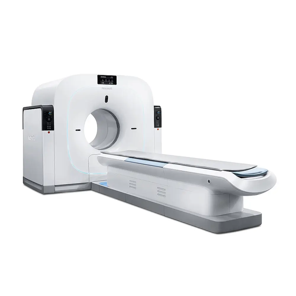
NeuSight PET/CT 64 Gantry NeuSight PET/CT 64 gantry consists of CT gantry and PET gantry PET Gantry Detector system Module PCB assy DAM assy Rod system Left electric cabinet Right electric cabinet
CT Gantry • High voltage generator • Tube • Detector :32 Rows(64-slice) PET Gantry Specification • Number of crystals : 17424 • Crystal size:4.7×4.7×30mm • Crystal material:BGO • Number of rings: 33; • Number of PMTs:576 • Ring diameter:856mm • Patient Port:72cm • Transverse FOV:70cm • Axial FOV:16.6cm • Rod type:68Ge PET Performance Index • System Sensiticity:7.0cps/kBq • Transverse resolution @1cm,mm :4.7;2.7(PDR) • Transverse resolution@10cm,mm :5.1;3.0(PDR) • Axial spatial resolution @1cm :4.8 • Axial spatial resolution @10cm :5.1 • Peak noise equivalent count rate :40kcps • Scatter fraction :45% CT Gantry Specification • Generator output :50kW • Output Voltage: 60-140KV • mA selections:30~420mA • Heat load :5.0MHU • Focal spot:0.6 mm x 1.2 mm (small) | 1.1 mm x 1.2 mm (large) • Number of detector row:32row(64-slice) • Number of detectors:672×32 • Detector material: Rare earth ceramic CT Performance Index • Spatial Resolution:17lp/cm@cut-off • Density resolution: 4.0 mm @ 0.3%@≤20mGy • SNR:≤0.35% • Range of CT Number: -1024 - +3072 Support CT number extension -10240~30720 • Number of slices :64 • Low Contrast Resolution:4.0 mm @ 0.3%@≤20mGy The main function and performance of NeuSight PET/CT 64 Tumor • Role in Oncology • Quantification:SUV • Differentiate benign from malignant disease • Staging of disease • Treatment response • Radiotherapy treatment planning Cardiovascular • Role in Cardiology • Myocardial perfusion and viability • Myocardial glucose metabolism • Myocardial receptor studies • Blood volume, wall thickness, wall motion Nerve system • Role in Neurology • Epileptic foci localization • Assessment of Alzheimer's disease and other forms of dementia • Assessment of Parkinson's disease and other movement disorders • Research of neuroreceptor mapping Health screening • The health screening of the high risk group with malignant tumor by PET/CT may facilitate the early discovery of malignant tumor. • The detection rate of malignant tumor of the normal group through routine health examination is 0. 3% ~0. 5%, while the detection rate of malignant tumor of the normal group through PET/CT is up to 2.2%. Meanwhile, most tumors were found at an earlier stage, so they have the opportunity for operation treatment. PET/CT Performance Parameters • NeuSight PET/CT 64 Image registration precision:≤3mm |
|
| 9 | Couch
• Vertical motion range:600-1030mm(Veetol) • Horizontal travel≥2570mm • Repeated Position Accurac≤0.25mm • Maximum patient weight :227kg • Adjustable scan height |
| 10 | Couch Cushion |
| 11 | Console Computer |
| 12 | PET Reconstruction Computer |
| 13 | CT Reconstruction Computer |
| 14 | Screen Display |
| 15 | PET/CT BOX
The PET/CT BOX can control the movement of the scan couch, the voice system and the scan. The PET/CT BOX mainly contains the following function buttons: |
| 16 | Computer Table
Tailor designed table for console cabinet. |
| 17 | NMS head supporter assy
To support patients’ head during scan. Extend couch scannable range. |
| 18 | Head supporter cushion |
| 19 | MMVA test tooling |
| 20 | PET calibration phantom |
| 21 | Water Phantom bracket |
| 22 | Water phantom bracket connection assy |
| 23 | QA Phantom |
| 24 | 7-10 Inch tower phantom |
| 25 | Nonlinear calibration phantom assy |
| 26 | Anchor template |
| 27 | Tool Box
A set of tools for daily maintenance to make sure the system to work with best status. |
| 28 | PET Gantry Separation Tool |
| 29 | 720 Patient Port of Rod Source System |
| 30 | Gantry Cover ABS Assy |
| 31 | Accompanying documents |
| AVW Post-processing Software & Advanced Algorithm | |
| 32 | NeuSight PET/CT AVW Operating System |
| 33 | NeuSight PET/CT AVW Software |
| The advanced application workstation is used for PET/CT image postprocessing, including the hardware and the software.
The hardware consists of the computer, monitor and network cable. The software consists of the AVW operating system and AVW software, which includes the following function: 2D display, advanced visualization, printing and reporting system. Surface Shaded Display Three-dimensional display of surfaces with different density values l Soft tissue l Bone l Contrast-enhanced vessels Multi-Planar Reconstruction(MPR) Variable slice thickness and distance with default values; Viewing perspectives l Sagittal l Coronal l Oblique l Double oblique Freehand (curvilinear) Print and Report System |
|
| 34 | Tumor Management
• Compare, analyze, and track tumor progression with up to four sequential PET/CT studies • SUV-based semi -automatic tumor segmentation • Measurement of changes in tumor volume and metabolic activity |
| 35 | Nerve Application
• Coregistration of PET or CT data with a reference template (The Talairach atlas). • Quantitative analysis of brain images in both Talairach and patient space. • Comparison of functional data with sequential brain images on the same patient. |
| 36 | Cardiac Fusion Software
• Analysis of 3D CT angiographic cardiac images/data providing a number of display, measurements and batch filming/archive features to study user-selected vessels. • Provide capability to visualize reformatted PET/CT perfusion and viability data. • Provide different ways of visualizing CT anatomy fused with PET functional information. |
| 37 | Cardiac acquisition system
The cardiac acquisition system could assist the clinician in using automatic tracking software of CT contrast medium to reconstruct the data based on the time phase of cardiac cycle, which could find optimal diastole and systole and perform multi-phase time phase reconstruction. The system acquires data by means of prospective and retrospective scan modes. The cardiac acquisition system helps the clinician to acquire PET multi-temporal data during cardiac cycle and edit ECG-gating parameter to conduct offline multi-temporal reconstruction. Moreover, you can choose the CT data at a particular phase as attenuation correction data to conduct offline PET image reconstruction. |
| 38 | Dynamic Viewer Software
Dynamic View software is available in console and/or advanced application workstation. |
| 39 | PDR
Point Diffusion Restoration Use PSF (Point Spread Function) technology to improve image resolution and reduce image noise. |
| 40 | Large FOV
PET reconstructed transaxial Display Field of View (DFOV) of up to 70cm with CT based PET attenuation correction and up to 70cm CT DFOV Wide FOV reconstruction |
| 41 | Free Match
Mis-registered PET and CT attenuation correction data due to organ motion may be re-aligned and reconstructed again to try and recover proper PET attenuation correction to help avoid CT AC re-scans . |
| 42 | Low Noise Reconstruction Technology
Add correction factor to iterative reconstruction and lower the noise caused by correction factor. Under the same image acquisition condition, this will improve image SNR compared with conventional reconstruction. |
| 43 | TIR
Target Iterative Reconstruction The target iterative reconstruction aims at small object target in the image to restart iterative reconstruction and realize the reconstruction of high pixel image. |
| 44 | Low dose CTAC
Reduce CT tube current, optimizing CT tube voltage to reduce dose of CT scan which is used for PET attenuation correction. |
| 45 | PVC
Partial Volume Correct Correct partial volume of the image. |
| 46 | ClearView
ClearView is an advanced iterative algorithm for double spaces, one is for original raw data space and another is for image space. With three levels of Slight, Standard and Ultra we can get perfect image quality with lower dose. |
| 47 | WLS
Weighted Least Square can effectively restrain noise image. |
| AVW Post-processing Hardware | |
| 48 | BeiTai Monitor HL1916S
Dual screen avaliable, both scanning and diagnostic can be working at the same time, Largely improve work efficiency. |
| 49 | DELL 5810 |
| 50 | BW Workstation reticle (10m) |
| 51 | UPS
UPS (Uninterruptible power supply) guarantees that the PET gantry, the patient table and the console could work for a period of time after a power failure so that the customer can save the data. No work would be affected or no data would be lost because of the power failure. |
| 52 | Isolation transformer
In order to meet the power supply voltage standards of different countries, the input voltage would be converted into 380V/220V to ensure the normal operation of the PET/CT. |
| 53-56 | UPS output cable(PET)
Isolation Transformer input cable Isolation Transformer output cable(CT) Isolation Transformer output cable(UPS) Cable used to connect the power distribution system with the gantry. |
| 57 | Brachiocephalic pad |
| 58 | Cervical vertebra cushion
Position cushion used for Cervical vertebra study. |
| 59 | Knee pads
Position cushion used for Knee Joint study. |
| 60 | Arm Support
For supporting patient's arms during intravenous injections. |
| 61-64 | Belt(Belt1-4)
Fasten patient on the couch to avoid motion artifact. |
| 65 | Table Stick
For moving the couch. |
| 66-68 | Cephalic pads
The cephalic pads consist of three sizes: large-sized, medium-sized and small-sized. |
| 69-70 | Coronary scanning head support Assy
The assy consists of coronary scanning head support and coronary scanning cushion. |
| 71-72 | Topo scanning head support Assy(includes head rest cushion.)
A set of cushions for better patient comfort while doing topo scanning. |
| 73-74 | Mindray iPM8 patient monitor and the cable
For international market, the default option is Mindray iPM8 patient monitor, |
| Tooling Bag | |
| 75 | patient table transport wheel kit |
| 76 | CT gantry transportation Dolly |
| 77 | PET gantry transportation connector |
| 78 | PET/CT patient table handling tool |
| 79 | Laser Leveler |
| 80 | Couch Fixed Target |
| 81 | CT Fixed Target |
| 82 | PET Fixed Target |
| 83 | Ultra-thin type hydraulic jack FPY-5 |
| 84 | Manual hydraulic pump CP-180 |


